
Cardiac catheter – Examination
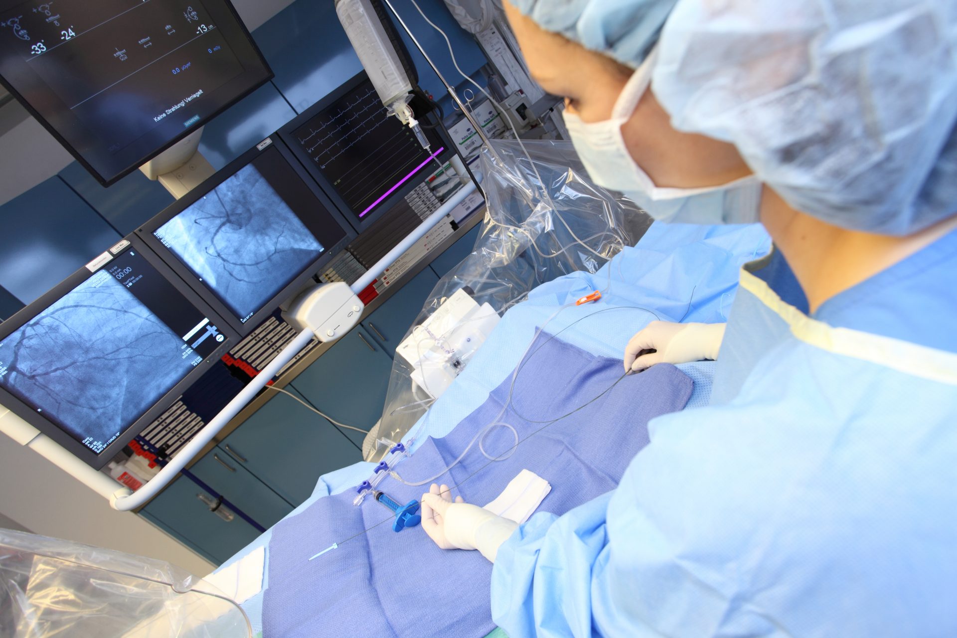
With regard to the cardiac catheter examination, a very thin tube is inserted through a blood vessel in the groin, elbow or wrist, which is used to access different sites in the heart or coronary vessels depending on the subject of the examination. The target zone determines the choice of access point.
A right-heart catheter should be used to examine the right ventricle and left-heart catheter for the left ventricle. Cardiac catheter examinations of the right ventricle are less common than cardiac catheter examinations of the left ventricle. This examination is frequently carried out to show and widen the coronary vessels in the context of a coronary angiography.
Cardiac catheter examinations provide information on the state of the examined sites, blood flow, pressure, oxygen saturation and temperature in the vessels, as well as electrical activity. A cardiac catheter examination is often immediately coupled with other treatments. Common accompanying treatments are balloon dilatations to address the blocking or constriction of coronary vessels, the implantation of a stent to support the blood vessels, the correction of congenital heart defects, such as valvular stenosis or septum defects, and even the implantation of heart valves without a massive surgical procedure.
Cardiac catheter examinations are normally carried out under local anesthetic. Apart from a feeling of warmth when the contrast agent is introduced, the patient is not aware of much regarding such an operation. The puncture site is closed with a pressure bandage once the treatment is complete.