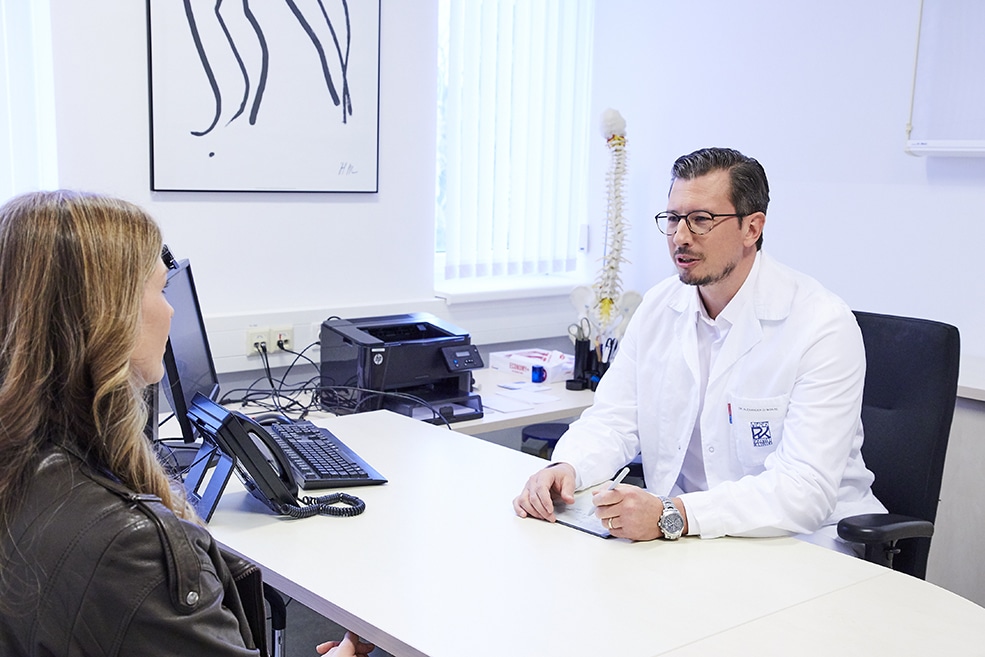
This website uses cookies to give you the best possible service. By using this website, you agree to the use of cookies. Detailed information can be found in our privacy policy.
OK
divider image

Check-up, Prevention

and Diagnostics Clarification

Benefits of Frequent Medical Check-ups & Advantages of Early Detection

Early detection can save your life. How? Well, besides leading a healthy lifestyle, regular check-ups are the best way to ensure receiving treatment for serious diseases that may or may not have any profound markers or evident, external symptoms. In which case, such knowledge calls for a precautionary check-up to enable detection and high chances of recovery.

Believe it or not, medical check-ups play quite a role in life expectancy. When Austria first introduced the comprehensive preventive medical check-up in 1974, the average life expectancy for women was around 75 years and 67 years for men. Today, women live seven years longer and men live eight years longer on average.

Why visit Wiener Privatklinik for a preventive medical check-up?

Wiener Privatklinik offers check-up packages that are tailored to your living conditions with a high level of competence and with the intention of ensuring patients are comfortable while receiving outstanding service. All preventive medical check-ups are carried out by utilizing the latest medical equipment under the direction of highly qualified medical specialists. Your examination program can be carried out on either an outpatient or on an inpatient basis – all in the comfort of our elegant hospital rooms with 5-star hotel amenities.

Which examinations are available at Wiener Privatklinik?

WPK offers an array of comprehensive examinations. The selection of preventive medical check-ups and the intervals between sections of your individual check-up program is determined by age, gender, lifestyle, any previous condition, and family history.

Ophthalmological examinations

Even if you have perfect eyesight, it is highly recommended that you have an eye check-up at least every two years. The concept of early detection and treatment of eye diseases applies to people of all ages. It should be noted, however, that eye examinations do not only serve as visual acuity tests and necessary optometry examinations. The condition of your eyes informs the specialists about more than you may think, such as the condition of your veins and arteries more-so than any other organ. Several metabolic disorders affect your blood vessels and signs of eye diseases, such as glaucoma, high blood pressure, and diabetes are often detected at an early stage by way of eye examinations.

What happens during an eye examination?

All eye examinations begin with standard questions concerning your medical history, any complaints, and known diseases, following which an eye test is in order.

During the eye test, visual acuity is determined with regard to varying ability in terms of nearsightedness and farsightedness. This visual acuity test, also known as a vision test, is initially carried out without corrective lenses and then with corrective lenses. If necessary, the visual acuity test is followed by thorough examinations of visual function, e.g. colour vision tests, measurement of the field of vision, eye movement, and the position and the light reaction of the pupils.

The ophthalmologist then uses a slit lamp to examine the anterior chambers of the eye, which include the lids, conjunctiva, cornea, anterior chamber, iris, and lens. The doctor will usually take this opportunity to measure the interocular pressure.

The patient is then given eye drops to dilate the pupils in order to ensure that the condition of the ocular fundus and lens can be examined with a magnifying glass.
Diseases that can be detected during such a thorough eye examination include:

  • Underlying diseases behind frequent headaches, e.g. frontal sinusitis, tumors, glaucoma (high interocular pressure), or functional disorders of the eye muscles.
  • High blood pressure, which can be revealed by irregularities or constrictions of the blood vessels in the eye.
  • Diabetes in which the first signs can include a change in quality of sight, which can be caused by small round blood flow regions or deposits on the retina.
  • Risk of stroke, which can be discovered through characteristic constrictions of the retinal vessels.
  • Elevated cholesterol levels, which can have a profound effect on the eyes.
  • Multiple sclerosis (MS), which can become noticeable from early signs, such as double vision, limitations of the field of vision, and involuntary or rapid eye movements (REM).

Gynaecological examination

The fact that women are at a high risk of breast cancer and cervical cancer should be two convincing reasons scheduling regular gynecological check-ups. Both cancers are now a much less frightening prospect than was the case in past decades and a major difference between then and now is the fact that these cancers can be treated with high chances of going into remission when detected at an early stage.

The intervals at which you should undergo a gynecological examination differ from individual to individual, and they depend on factors such as age, family history, etc. You are guaranteed to receive the appropriate information needed during an initial examination at the Vienna Private Hospital.

What happens during a gynecological examination?

The main tests run during gynecological preventive medical check-ups are that of a mammography and a pap smear.

A mammography is an X-ray image of the breast in which the breast is placed between two X-ray Plexiglas plates. The mammography illustrates the internal structure of the breast in black and white contrasts and it makes even the smallest of changes visible. In case of doubt or abnormal findings, a breast ultrasound examination or MRI may be required. During the ultrasound examination, a contact gel is applied to the breast and it is then examined with a transducer. The transducer sends signals through the breast, which return as “echoes” within fractions of a second, and then appear on a monitor in the form of a black and white image.

The magnetic resonance imaging (MRI) serves to clarify suspicious or unclear mammography or sonographic results. During the analysis of the results, layered images of the breast are generated, which allow for distinguishing between tumor formations and healthy tissue. Meanwhile, the pap smear of the cervix plays a pivotal role in the early detection of cervical cancer. Cervical cancer predominantly occurs to women between the ages of 25 and 55. Moreover, it affects six out of 10,000 women within the aforementioned age range. Since the introduction of the pap smear test in gynecological practices, the mortality rate for cervical cancer has been reduced by two-thirds.

During the pap smear test, the cells of the cervix and the outer area of the uterine opening are extracted with a small brush or cotton swab and taken to the laboratory for analysis. It should be noted that an abnormal finding is no reason for panic. Slight to moderate cell changes are very common in young women under the age of 30. They do not necessarily indicate cancer. However, they can indicate an increased risk, which requires further check-ups at shorter intervals.

Dermatological examination

Having a dermatological medical check-up annually is certainly an advisable preventative measure against skin cancer. During the examination, existing moles and all newly developed dermatological changes are examined, regardless of slow progression of growth and even if they do not hurt or itch.

Under unfavorable conditions, moles can degenerate into skin cancer (melanoma). It should be noted that around 25 % of melanomas develop from moles.

What happens during a dermatological examination?

The dermatological examination or dermatoscopy entails a thorough visual inspection of the skin by the specialist through the use of oils, which allow for transparency of the upper layers of skin and thus provides a clearer picture. During the examination, the dermatologist looks for symptoms pertaining to three different degrees of skin cancer, which include:

  • Melanoma: a malignant tumor, which develops from the pigment-producing skin cells. Melanoma can be caused by high levels of UV radiation exposure. Individuals with fair skin, moles, and a family history of melanoma or various skin cancers have a particularly higher risk of developing melanoma. Melanoma can only be treated effectively when detected at an early stage.
  • Cutaneous squamous cell carcinoma: a malignant tumor, which often affects people with light-sensitive skin, who are exposed to an increased level of UV radiation for years and have weaker immune systems as a result.
  • Basal cell carcinoma: a common, yet less dangerous and more curable skin tumor.

Cardiovascular examination

Your heart is the untiring motor of your circulatory system and as such, it beats more than 100,000 times per day. It’s imperative that it functions without fault in order to ensure your safety and a greater quality of life. Regular cardiovascular examinations are highly recommended, alongside leading a healthy lifestyle in order to minimize your risk of disease and infarction.

What happens during a cardiovascular examination?

The range of methods employed in detecting a heart disease and issues in the circulatory system are quite remarkable. The initial information deduced from a cardiovascular examination is provided by a pulse and blood pressure measurement and auscultation with a stethoscope. In order to more accurately assess the condition of your heart and your coronary vessels, further examinations are often required. These include:

  • Resting ECG: an ECG records the electrical activity of the heart. During an ECG, small electrodes are attached to the upper body, which then send electrical pulses from the heart to an ECG device. The ECG detects cardiac arrhythmia, provides indications of previous myocardial infarction(s), and can also reveal an acute cardiac infarction.
  • A deeper insight can be provided by way of an exercise ECG or ergometry, which employs a bicycle ergometer or treadmill. During this examination, the function and performance of the heart and circulatory system are examined during physical exercise. Ergometry enables the assessment of blood pressure behavior, oxygen supply to the heart, signs of cardiac arrhythmia during exercise, cardiac weaknesses, and certain valvular heart diseases. The exercise ECG is much more informative than a resting ECG alone. Moreover, alongside the results from a blood pressure measurement, it is also the most informative general circulatory system examination. However, in case of suspicion, it will usually be accompanied by other examinations, such as a long-term ECG with a portable device. The long-term ECG with a portable device can record cardiac function during a period of at least 24 hours.
  • Echocardiogram: an echocardiogram is an ultrasound examination of the heart. An echocardiogram provides a clearer picture of the movement and structure of the heart. Moreover, it can reveal valvular defects and abnormal progression of heart contractions. However, abnormal changes in the coronary vessels cannot be seen through the use of an echocardiogram. Abnormalities in coronary vessels are typically diagnosed by means of angiography.
  • Angiography: During an angiography, a catheter is inserted into an artery in the groin – during which the patient is put under local anesthesia – and pushed up to the heart and coronary vessels. A contrast agent is then injected into the heart via the catheter, so that the coronary vessels can then be assessed by means of X-ray images. The existence, number, and precise location of the coronary vessels can clearly be seen on the X-ray film.

Respiratory examination

On average, humans inhale and exhale 20,000 liters of oxygen per day. As such, so long as the lungs are functioning without any impairments, the body is provided with a vital source of oxygen and the chance of inhaling poisonous carbon dioxide is greatly eliminated. Therefore, respiratory examinations are an important part of each comprehensive preventive medical check-up. The comprehensiveness and thoroughness of the examination depends on the individual condition of the patient.

What happens during a respiratory examination?

The specialists initially examine the patient for the external symptoms of lung disease, e.g. blue lips and fingers, changes of the fingernails, deformations of the chest, changes in breathing, coughing, etc. Specialists then feel whether the breathing movement of the chest is symmetrical, alongside checking on the thyroid gland, trachea, and lymph nodes by pressing or tapping. Breathing sounds and patterns are then checked with a stethoscope. This provides the specialist with an inside look as to what possible diseases of the lungs and pleura may be present.

In addition to examinations of the blood and saliva, deeper insights can be provided by lung function examinations, such as:

  • Spirometry / ergometry is used for determining the maximum breathing capacity and for diagnosing a constriction of the airways or reduced expansivity of the respiratory organs. During the examination, the patient must breathe into a mouthpiece whilst the nose is plugged.
  • Peak-flow measurement: The peak flow monitors the development of certain lung diseases, such as asthma whilst the patient is exhaling, so that deterioration can be counteracted as early as possible.
  • Body Plethysmography: In addition to the values provided by spirometry, body plethysmography plays a large role in determining the amount of gas volumes in the chest and resistance in the airways. Moreover, it assesses the conditions of two other parameters of respiratory function, which are liable to change in the presence of disease.
  • Thorax CT: The thorax CT (Computed Tomography) creates two-dimensional images of organs and tissue layer by layer, which can be combined to form three-dimensional images to gain a spatial impression of the organs. Tissue, organs, and changes can be displayed free of superimposition in CT images and thus, they are much more efficient in detecting and illustrating abnormalities in comparison to conventional X-ray images and machines.

Gastrointestinal tract examination

Preventive medical check-ups of the gastrointestinal tract serve to detect and treat cancers at an early stage. The risk and the importance of this particular examinations increases with age. Gastrointestinal examinations for bowel cancer prevention should be carried out routinely every 5-7 years once an individual passes the age of 50. Regardless of whether a disease is present at any given time, a comprehensive examination is advisable for the sake of ensuring a long-lasting life free of gastrointestinal problems.

What happens during an examination of the gastrointestinal tract?

The gastroscopy, gastric examination, and colonoscopy are the most informative methods with regard to approaching the diagnosis of gastrointestinal diseases. A thin, flexible endoscope with a video chip at the tip allows the doctor to assess the condition of the mucous membrane, painlessly extract tissue samples, and, if necessary, carry out simple procedures, such as polyp removal and homeostasis, during the examination.

Gastric examinations typically take around 10 to 15 minutes. This examination enables specialists to assess the state of the esophagus, stomach, and duodenum. Tissue samples are extracted for the sake of assessing changes in the mucous membrane and detecting any bacterial infestations of the mucous membrane.

Colonoscopies typically take around 20 to 30 minutes and is painless. This examination enables specialists to get a clear view of the entire colon and the entrance of the small intestine. With regard to bowel cancer prevention, polyps, which can be detected during this intestinal examination, are immediately removed. Tissue samples are extracted for the clarification of diarrhea and the existence of enteritis.

Orthopaedic examination

Spinal pain and joint pain can occur for an array of reasons – some of which can severely affect a person’s quality of life. Some pains in one’s spine and/or joints can even hint at life-threatening diseases, such as an aortic aneurysm or shingles. In order to effectively pinpoint causes of orthopedic pain, a thorough examination is required. The orthopedic examination serves to clarify causes behind occurrence, to prevent and watch out for dangerous diseases or nerve damage, and to pave the way for therapy, which can rejuvenate one’s range of mobility and aid one in returning to as pain-free of a life as possible in terms of more severe pains.

What happens during an orthopedic examination?

The orthopedic examination begins with a visual inspection of the vertebral column to investigate whether the patient has a hollow back, a hunched back, a flat back, etc. Furthermore, whether the shoulder blades and waist symmetrical is investigated, alongside checking if the vertebral column and pelvis are straight. Smaller diversions from symmetry are not cause for concern, but larger diversions can be an indication of a disease, such as scoliosis. Postural defects, as a result of scoliosis or possibly other abnormalities, can cause chronic back pains and are quite easy to detect by checking the gait when the patient takes a few steps back and forth.

In the next examination, the specialist examines the vertebral column and back muscles for differences and tensions. The mobility of the vertebral column is then examined through various test exercises.

If these tests cannot be done during orthopedic examination due to pain, a neurological examination is required to rule out possible nerve damage. During the neurological examination, the specialist will test different reflexes amongst other things in order to assess the presence of nerve damage or rule it out as. If necessary, an MRI can then take place to provide the specialist with a look into deeper structures.

Metabolism examination

An adequate metabolism examination is highly recommended due to the fact that the various cycles that the body goes through, day after day, shapes our lives and heavily determine our overall well-being. Metabolic disorders, metabolic diseases, and food intolerances can have drastic effects on one’s quality of life. A metabolism examination allows for the assessing the state of the patient’s hormones, metabolism and tolerances, and energy consumption whilst resting and whilst partaking in physical exercise. Nutritional changes and therapeutic recommendations can be configured to work around individual requirements.

What happens during a metabolism examination?

There are many metabolic disorders – many of which can affect various organs in the body, such as the heart, thyroid gland, liver, and brain amongst others. Said metabolic disorders range from common diseases, such as gout, diabetes mellitus, and hypothyroidism/hyperthyroidism, to various allergies.

Symptoms of many metabolic disorders include strong physical discomfort, sleep disorders (e.g. sleep apnea, etc.), mood swings, drastic changes in body weight, abnormal skin changes, and hair loss amongst others.

The presence of such symptoms is cause for concern and calls for a doctor’s visit. The urgency of the situation is made clearer through finding out about the patient’s blood count and specifying the severity of the symptoms.

Metabolic diseases are often characterized by an enzyme deficiency or hormone deficiency. With regard to some metabolic disorders, some metabolism products accumulate in the body whilst others may be stored or excreted. However, transport defects due to certain substances can also occur, i.e. a rise in blood sugar due to diabetes mellitus as a result of a lack of insulin.

The prevalence of allergies and food intolerances calls for concern, as they are often the cause for chronic gastrointestinal complaints, and they are also involved in the development of numerous unclear complaints, such as chronic tiredness, energy loss, sleep disorders, susceptibility to infection, headaches/migraines, rheumatic complaints, chronic bronchitis, the inflammation of skin, mucosal membranes and joints, and cardiovascular problems amongst others.

Specialized screenings, such as EAT, alongside special laboratory diagnostics, are often used to discover reasons behind allergies and intolerances.

Urological examination

Men over the age of 45 should seriously consider having an urological examination done as a way in the risk of prostate cancer can be monitored. Malignant changes to the prostate are the second most common cause of death in men in terms of cancer. An annual urological check-up ensures that changes to the prostate are detected and treated long before the occurrence of noticeable and far more serious symptoms can arise.

What happens during an urological examination?

The physical examination begins after a detailed interview takes place in which medical history is to be discussed. In urology, the kidneys and their environment are examined by way of employing an ultrasound examination whilst the bladder is examined through palpation. Meanwhile, the genitals are examined for skin changes and swelling. The prostate is then examined by way of a rectal examination.

In order for the doctor to gain a more detailed picture of the medical condition of the patient, it is usually necessary to follow-up the physical examination with further tests. Specifically, the examination of the urine, blood, and neurological examinations may be advisable.

Our hospital combines excellent specialists, medical expertise, and state-of-the-art facilities for all diagnostic steps:

Blood test

A blood test is defined as the extraction of a small amount – approximately 2 to 50 milliliters – of venous or arterial blood, which provides an in depth look into the blood’s contents and solid particles by utilizing different laboratory methods. The results include a diagnostically relevant blood count, which indicates the state of one’s medical condition and hints at any diseases or risks of disease, amongst other revealing information (e.g. iron deficiency = anemia, etc.).

When is it advisable to have a blood test done?

There are several manners in which one can have a blood test done. A single blood sample can provide up to 40 different pieces of information on the medical condition of a patient. Therefore, blood tests are standard for almost all preventive medical check-ups in which blood counts specifically are a fixed component of the general clinical examination for preoperative preparations, inflammations, tumors, infections, coagulation disorders, anemia, and blood diseases.

What occurs during a blood test?

Blood samples can be arterial or venous. Venous blood is used for the majority of blood tests. However, arterial or capillary blood samples may be necessary for individual examinations.

Venous blood samples are normally taken from the inside of the patient’s elbow. The patient should be sitting down or lying down at the time. In order to ensure the vein is fully visible, the blood flow is constricted for a short period of time at the upper arm by means of a tourniquet or a cuff. The puncture site is cleansed with a disinfectant, following which the vein is punctured with a needle and the amount of blood required is extracted with a syringe or a vacuum blood collection tube. A gauze pad or plaster is then applied onto the puncture site for around a minute so that the bleeding stops. During that minute, the elbow should not be bent, so that the vein is not reopened and a bruise is not caused.

Blood test – are there alternatives?

Blood tests are painless and there are no accompanied risks. As such, there is no better alternative. If minuscule amounts of blood are required, e.g. for blood glucose level, then the fingertip or the earlobe can be pierced with a small lancet and a few drops of blood can then be squeezed out.

Computed tomography

Computed tomography (CT) should actually be called X-ray computed tomography, as CT is a computer-aided X-ray examination. In contrast to the “classic” X-ray, which only captures a two-dimensional image on the X-ray film, computed tomography provides cross-sectional images of the body from which three-dimensional images are generated. The three-dimensional images not only serve to detect various diseases, but also determine the position and extent of abnormal changes.

Today, computed tomography is a standard piece of equipment to have at clinics and doctor’s offices. It is used in the diagnosis process for various diseases and for monitoring the progress of treatments. For instance, a CT can track the status of a suspected tumor, stroke, slipped disc, bone fracture, examinations of the coronary vessels, and early detection of bowel cancer amongst others. X-ray images of the chest are frequently supplemented by a thorax computed tomography (Thorax CT).

What occurs during a CT?

The patient is pushed through the device, whilst the X-ray tube continually rotates around the patient. The examination is normally carried out in the supine position on the CT table. It typically takes anywhere between a few seconds and 15 minutes at most. Cross-sectional images in any desired region of the X-rayed body can be generated from the recorded data. For example, an image of the chest is taken with a Thorax CT. During or after the Thorax CT, the computer can convert the single cuts into a three-dimensional image.
For certain procedures, contrast agents may be used. Contrast agents are either administered to the patient via the vein or the mouth. Moreover, employing contrast agents can enhance the resolution or quality of the computed tomography.

Computed tomography – are there alternatives?

In comparison to a CT, ultrasounds (sonography) and magnetic resonance imaging (MRI) have the advantage that they do not expose the patient to X-rays. Today, some examinations, which have long been the domain of computed tomography, are carried out by way of an MRI instead. Each patient must make a decision as to which imaging method is best suited to their needs. It’s important to remember that the CT, MRI, classic X-ray, and ultrasound are not interchangeable. Computed tomography is often used when “more gentle” methods have come back empty.

Colonoscopy

Colonoscopy – how?

The tool used in a colonoscopy is the endoscope (coloscope), a flexible tube which adapts to the bends in the colon. There is a video chip at the tip, which transmits the recorded images to a monitor (video endoscopy). There are additional channels in the tube, such as cleaning and suction devices and channels for inserting surgical instruments. Smaller interventions can also be carried out as part of the endoscopy/rectoscopy, such as homeostasis, tissue sampling (biopsy) or polyp removal. In order to show the entire colon, the coloscope has a length of approximately 1.3 meters.
The examination is not very unpleasant and risk-free to a large extent. For those with sensitive natures, the colonoscopy can be carried out after administering sedatives or light anesthesia.

Colonoscopy – are there alternatives?

The virtual or CT coloscopy is a possible alternative to the endoscopic examination. Two and three-dimensional images of the colon are generated by means of high-resolution multi slice CT devices and special computer programs and examined for changes, such polyps or tumors. The colonoscopy and CT coloscopy are able to detect polyps with the same degree of precision. However, the conventional coloscopy offers the major advantage that such polyps can be removed in the same examination step. Patients whose polyps are detected during a CT coloscopy often require an additional colonoscopy for polyp removal. The radiation exposure from a CT examination can be seen as a disadvantage, even though it is low. The CT coloscopy is mainly used in case of severe constrictions of the colon, which cannot be passed by an endoscope. It can also be considered as an alternative in case of other contraindications for a colonoscopy.

ECG – Echocardiography

During the electrocardiography (ECG), the electrical activity of the heart is recorded in the form of a curve on an electrocardiogram. In the heart, there is a weak current with a voltage of around one thousandth of a Volt per beat. These electrical pulses are deduced using electrodes (metal platelets). For this purpose, the electrodes are applied to the arms, legs and chest. An ECG device amplifies these pulses and presents them as a curve on a screen or prints them on a strip of paper.
Thus, electrocardiography is the recording of the course of cardiac excitation.

ECG – when?

With the electrocardiogram, the doctor examines

  • Cardiac rhythm and heart rate
  • Disorders of the ectopic rhythm, propagation and regression in the impulse conduction system and in the cardiac muscles
  • The position of the heart in the chest

The following can be detected by curve changes in the ECG:

  • Cardiac infarction
  • Coronary vessel diseases
  • Overdosage of certain medications
  • Lack or abundance of certain minerals and their influence on impulse conduction.
  • Atrial fibrillation or atrial flutter and ventricular fibrillation or ventricular flutter
  • heart beat that is too slow (bradycardia) in diseases of the sinus node or AV node
  • Inflammation of the myocardium (myocarditis)
  • Inflammation of the pericardium (pericarditis)

The treatment and progress of different heart diseases are often monitored by means of ECG.

ECG – how?

During a resting ECG, the patient lies down in a relaxed physical state. The measuring points are on the arms, legs and the chest. They are connected to the ECG device which then measures the electrical activity of the heart. After the examination, the doctor removes the measuring electrodes and evaluates the ECG.

During an exercise ECG, the patient must be active on either a treadmill or a room bicycle. This is useful:

  • if there is a suspicion of coronary vessel diseases
  • if there is a suspicion of exercise hypertension (abnormal increase in blood pressure during exercise)
  • for the clarification of physical fitness after cardiac infarction or following cardiac surgery
  • for the assessment of heart rhythm disorders during exercise
  • for the assessment of medicinal treatment of coronary vessel diseases or increased blood pressure

A long-term ECG records the electrical activity of the heart over a period of at least 24 hours. In this case, the ECG device is particularly small so that, for example, it can be carried on the belt.
During exercise tests, cardiorespiratory fitness and changes in cardiovascular function are examined and assessed for different diseases, e.g. high blood pressure, bradycardia, chronotropic incompetence, heart failure, valvular heart disease, cardiac arrhythmia, coronary heart disease (CHD) and following heart surgery.

Ergometry

Ergometry – how?

You should wear comfortable sportswear and trainers for the exercise test on the bicycle ergometer or the treadmill. At the start of the examination, a resting ECG is recorded and the resting blood pressure is measured. For this purpose, several electrodes are attached to the skin on the chest and back and a blood pressure cuff is applied to the left upper arm. The ECG recordings during physical exercise are then repeated at regular intervals.
The physical exercise is either on a bicycle or a treadmill. The resistance of the bicycle ergometer or the speed of the treadmill is increased every two minutes to increase the level of exercise in a controlled manner. The examination is carried out until the exercise limit, unless certain measurement values (such as blood pressure or heart rate) exceed the determined limit values or if heart complaints or breathing difficulties occur. Once the exercise limit is reached, you should keep peddling or walking without effort for a few more minutes, so that complaints which occur a short period of time after exercise can be detected.

Ergometry – are there alternatives?

Ergometry is just one examination in a whole arsenal of cardiovascular examinations.
The worst side-effect for healthy patients is a slight muscular ache. With regard to patients with severe cardiac disease, the examining doctor must carefully consider whether there are any contraindications for the implementation of ergometry (e.g. acute coronary syndrome, myocarditis, serious

– if they are bleeding – they can also be treated immediately.

Further applications are the removal of polyps (benign mucosal proliferation), the removal of swallowed foreign bodies and the widening of constrictions in the oesophagus or the area of the pyloric orifice. For the latter, an inflatable balloon is pushed through the working channel of the endoscope.

Gastroscopy

Gastroscopy – how?

A special endoscope, the so-called gastroscope, is used for the gastric examination; a flexible tube which can be controlled and moved by the doctor. Inside, in addition to a working channel and an air/cleaning channel, there is a video lens which transmits images to a monitor.
The instruments are still smaller than a centimetre thin and they can be pushed into the stomach via the mouth and oesophagus without any problems.
During the examination, the patient lies on their left side. The gastroscope is then pushed through the mouth via the oesophagus into the stomach and also the duodenum if a diagnosis is also required there. In order to unfold the mucous membranes and thus for a better assessment, the stomach is inflated with air for a gastroscopy. The doctor then sees the inside of the upper digestive tract, piece by piece, via the integrated lens. If they detect changes, they can take mucous membrane samples with small forceps passed through the working channel, which are then examined in the laboratory.
The duration of a gastric examination is normally only a few minutes. Once it is complete, the doctor slowly pulls the device back and then straight out. The previously injected air is extracted to prevent or reduce excessive belching and bloating.
The method does not cause pain. However, the insertion of the gastroscope can cause an unpleasant gag reflex. Meanwhile, there are also devices which are pushed through the nose, which many patients perceive as more pleasant.

Gastroscopy – are there alternatives?

All in all, the gastroscopy is a very safe procedure, which has long become routine practice in hospitals and doctor’s offices. In many cases, there are no alternatives, as the Lipid profile

Lipid Profile

The lipid profile is part of the blood test in which the presence of HDL cholesterol, LDL cholesterol, total cholesterol, and triglycerides in the blood are determined with laboratory resources. This examination, which carried out regularly, plays a major role in the prevention of atherosclerosis and cardiovascular diseases.

Biochemistry has taught us a lot about the function of different blood lipids. Cholesterol, which is partly ingested with food and partly synthesised in the liver, is an essential component of cell membranes. Triglycerides primarily serve as sources of energy and storage substances. They also form the mostly undesired fat pads on the body and they represent a form of energy that can be continued during physical exercise. Lipoproteins, such as HDL and LDL, serve to transport the cholesterol in the blood. The basis for an assessment regarding the percentage of risk involved in terms of arteriosclerosis and thus, to ensure suitable recommendations ensue for a health-conscious lifestyle can come by way of investigating the distribution of cholesterol, HDL, LDL and triglycerides in the blood. This can be determined with a blood test.

When it is advisable to check on your lipid profile?

The lipid profile can be checked with simple blood test at the Wiener Privatklinik as part of preventive medical check-ups. It serves as a crucial decision-making factor regarding what nutritional and lifestyle recommendations the specialist will make for the sake of prevention of cardiovascular diseases.

Mammography

Mammography – are there alternatives?

The X-ray examination of the breast – the mammography – is the most important internationally recognised and standardised method for the early detection of breast cancer. Magnetic resonance imaging (MRI) is also used in exceptional cases.
The ultrasound examination (sonography) of the breast is another established method; however, it accuracy depends on the breast density. In contrast to the Mammography, it is not possible to detect so-called microcalcification, which can indicate a surface carcinoma in the mammary ducts in some cases.

MRI – Magnetic resonance imaging

MRI – Magnetic resonance imaging – Process, benefits and alternatives

MRI – what is involved?

Magnetic resonance imaging/MRI is an imaging method, with which organs and tissue can be shown in great detail and any changes can be assessed. The examination method also known as nuclear spin tomography uses magnetic fields and high-frequency electromagnetic waves instead of X-rays.

Cross-sectional images of the inner body can be produced using magnetic resonance imaging. Soft tissue, such as the brain and internal organs, show up high in contrast on an MRI image and differentiated so that even details less than a millimetre in size are discernible. In addition to this high resolution, another advantage of the method is that the images can be taken in any desired plane – i.e. not only crosswise through body, but also lengthwise or diagonally. The computer can then generate a three-dimensional image of the inner body from the cross-sectional images.

MRI – when?

Magnetic resonance imaging is an essential medical instrument both for the detection and exclusion of a disease and for the monitoring of therapies. With the exception of organs with a low water content, bones or the lungs, MRI provides highly precise images of almost

MRI – are there alternatives?

When bony structures or the lungs are examined, the classic X-ray and computed tomography (CT) are more suitable than MRI. In other respects, the procedure is often used as an alternative or supplement to other diagnostic methods when their limits are reached.
I.e. magnetic resonance imaging is usually only carried out if other techniques, such as ultrasound, X-ray and CT do not provide reliable results, or at least sufficiently reliable results. Accordingly, there are also no alternatives for many issues.

PSA test

“PSA” stands for “prostate-specific antigen.” PSA is a protein molecule, which is produced in the prostate. The concentration of PSA can be measured in the blood.

In general, malignant changes in the prostate gland can lead to an increased distribution of this protein molecule in the blood. Nowadays, attempts are made to use this in early detection. Today, more than 80 percent of all newly discovered prostate carcinomas are only detected by the PSA test. The difficulty here is that there is no clear lower limit from which prostate cancer can be excluded with certainty and that the increased PSA level has other, comparatively harmless causes in many cases, such as that of prostate inflammation.

When is it advisable to have a PSA test done?

A PSA test may be recommended as part of a prostate examination, particularly as part of cancer screening. However, the potential stress caused by a conspicuous PSA level should be openly discussed between doctor and patient.

How is a PSA test done?

The PSA test is included in the blood test and requires nothing more from the patient than a blood sample.

PSA test – are there alternatives?

The test is only part of a comprehensive preventive medical check-up. The fact that there still remains debate about its value among experts is due to it not being a standard facet of blood tests at clinics or hospitals worldwide. Therefore, people should obtain comprehensive information about the PSA test from their doctors in order to reach a joint decision on whether or not they wish to make use of the test. However, there is no doubt that the PSA level is essential for the diagnosis and monitoring of a previously detected prostate carcinoma.

X - ray examination

In many areas of medicine, X-ray is the fastest, most reliable and proven method of detecting injuries or diseases. During an X-ray examination, X-rays which are generated by an X-ray tube are sent through the body. These rays are then captured on the other side by means of X-ray film or digital storage media.
While bones and other tissue with a very high calcium content (such as gall or kidney stones) absorb a large part of the radiation and they become very visible in the X-ray images, soft tissue and hollow organs are very radiolucent and thus they are less visible. In order to improve clarity, a contrast-enhancing agent
(contrast agent) is used in certain cases.

X-ray examination – are there alternatives?

If you are pregnant or if a pregnancy cannot be excluded with certainty, the examining doctors will decide not to carry out an X-ray examination in most cases. In any case, a precise risk-benefit analysis will be carried out before you are exposed to radiation. Where appropriate, other potential examination methods will be used.

X-ray examination – when?

X-ray diagnostics are particularly well suited for representing the bony skeleton (i.e. detecting bone fractures or malpositions), but also showing lung diseases (such as also showing lung diseases (such as inflammations, tumours or fluid accumulations). To this day, mammography, the X-ray imaging of the mammary glands, is the method of first choice for the early detection of breast cancer. However, vessels (arteries, veins, lymphatic vessels), hollow organs (gastro-intestinal tract, kidneys, efferent urinary tract, gallbladder and biliary tract, tract systems), artificial cavities in the body (such as fistulas or abscesses) as well as organs and soft tissue can also be made visible and assessed in computed tomography using X-ray contrast agents.

X-ray examination – how?

During the X-ray examination, the body is exposed to a higher level of X-ray radiation than in everyday life, i.e. earth radiation or radiation from television sets, for a short period of time. Modern X-ray machines with short exposure times keep the radiation exposure extremely low. Particularly radiosensitive organs (such as the genitals) are shielded with lead aprons or screens to keep the radiation dose for the organs as low as possible. If a contrast agent is used for the examination, it is either injected into the bloodstream via a visible vein or artery by means of catheter or injection, or it enters the gastro-intestinal tract through drinking, depending on the case.

Two-dimensional images allow the doctor to gain a spatial view of the size, shape and structure of the investigated organs. Besides the normal structure of the liver, spleen, heart, kidneys and other organs, changes in blood vessels and soft tissue can also be shown.

Ultrasound examination

The ultrasound examination or sonography is the examination of organic tissue by means of ultrasound. Today, it is one of the most frequently used imaging processes in medicine, as many diseases can be examined and clarified faster, and thus with reduced costs and risks, through ultrasound examination. Furthermore, the treatment is completely harmless and free of side-effects.
During ultrasound examinations, sound waves with a frequency above the audible range of the human ear are sent through the body, reflected by structures and tissue and registered by a receiver. A computer creates an image from this information.

The flow rates of fluids in vessels and the heart can be converted into an image with special pulse forms.

Ultrasound examination – when?

Ultrasound examinations of the breast (mammasonography or breast ultrasound examination), the heart (cardiac ultrasound) and the important main arteries (carotid ultrasound) as well as examinations during pregnancy are just the most prominent examples for the wide range of applications of this examination method.

Ultrasound examination – how?

The ultrasound examination is painless and completely free of side-effects. The examining doctor touches the patient with a sensor and the image of the examined body region appears on a monitor.

Ultrasound examination – are there alternatives?

As no dangerous rays are released, the ultrasound examination is completely harmless. The real question is what are the alternatives to ultrasound when the method reaches its limits. Because not all organs are as well suited for ultrasound examination.

We are available to you at any time for any questions you may have and you can request counselling interviews with highly qualified doctors if you would like to obtain a second opinion before proceeding with an operation or therapy treatment.

We are happy to include existing documents, which you should please forward to us, in the diagnostic clarification.


Our office is always at your disposal for specific diagnostic examinations and consultations with renowned, highly qualified physicians. Especially if you want to get a competent second opinion before a surgery or a therapy.

We would be pleased to include existing documents in the diagnostic evaluation, which you should send to us in advance.

For further informations, please contact us at info@wpk.at.

We would be happy to make an individual offer for you.

Check UP Programs

Basic, Plus, Deluxe, KiDS

Basic Check

  • Medical consultation with a specialist
  • Advanced laboratory (blood, urine test)
  • Blood pressure control
  • Resting – EKG
  • Body fat / BMI
  • Ultrasound heart and carotid artery
  • Breakfast at the WPK Cafeteria
  • Final consultation

cost estimate on request

Health Check Plus

  • Medical consultation with a specialist
  • Advanced laboratory (blood, urine test)
  • Blood pressure control
  • Resting – EKG
  • Body fat / BMI
  • Ultrasound heart and carotid artery
  • Chest X-Ray
  • Ultrasound:
    • Abdomen
    • Kidneys
    • Pancreas
    • Liver
    • Thyroid
  • Breakfast at the WPK Cafeteria
  • Final consultation

cost estimate on request

Health Deluxe Woman

  • Medical consultation with a specialist
  • Advanced laboratory (blood, urine test)
  • Blood pressure control
  • Resting – EKG
  • Body fat / BMI
  • Ultrasound heart and carotid artery
  • Chest X-Ray
  • Ultrasound:
    •  Thyroid
    •  Abdomen
    •  Kidneys
    •  Pancreas
    •  Liver
    •  Thyroid
    • Breast
  • Special consultations:
    • Gynaecological consultation
    • Vaginal ultrasound
    • PAP smear test
    • Bacterial culture smear
    • Pulmonary function test
    • Dermatological examination and consultation
    • Eye examination and consultation
    • Mammography
    • Coloscopy and gastroscopy
    • Dietological consultation
  • Breakfast or lunch at the WPK café
  • Final consultation

cost estimate on request

Health Deluxe Man

  • Medical consultation with a specialist
  • Advanced laboratory (blood, urine test)
  • Blood pressure control
  • Resting – EKG
  • Body fat / BMI
  • Ultrasound heart and carotid artery
  • Chest X-Ray
  • Ultrasound:
    •  Thyroid
    •  Abdomen
    •  Kidneys
    •  Pancreas
    •  Liver
    • Thyroid
  • Special consultations:
    •  Urology
    • Eye examination and consultation
    • Pulmonary function test
    • Skin examination and consultation
    • Coloscopy and gastroscopy
    • Dietological consultation
  • Breakfast or lunch at the WPK café
  • Final consultation

cost estimate on request

Infant Check

3rd month of life

  • anamnesis with specialist
  • family anamnesis with specialist
  • resting – ECG
  • blood pressure
  • ultrasound:
    • heart
    • abdomen
    • head – brain
  • final talk

cost estimate on request

Kids Check

kids 2 – 6 years

  • anamnesis and physical examination with specialist
  • resting – ECG
  • blood pressure
  • extended laboratory
  • ultrasound:
    • heart
    • abdomen
    • transcranial Doppler
  • final talk

cost estimate on request

Adolescence Check

kids 14 – 18 years

  • anamnesis with specialist
  • Endocrinological assessment and physical development (hormonal status)
  • extended laboratory
  • blood pressure
  • resting – ECG
  • exercise -ECG
  • ultrasound:
    • carotis
    • heart
    • abdomen
    • transcranial Doppler
  • final talk

cost estimate on request

Add – Ons

Eyes

  • Eye examination and consultation

cost estimate on request

Gastroscopy / Coloscopy

  • Gastroscopy
  • Coloscopy
  • Final consultation

cost estimate on request

Gynecology

  • Gynecological consultation
  • Vaginal ultrasound
  • PAP smear test
  • Mammography
  • Breast ultrasound

cost estimate on request

Heart

  • Cardiological consultation
  • Ultrasound heart
  • Exercise ECG
  • Final consultation

cost estimate on request

ENT

  • ENT examination and consultation

cost estimate on request

Kids - Eyes

  • Ophthalmological examination and consultation

cost estimate on request

Kids - HNO

  • ENT examination and consultation

cost estimate on request

Kids - Hormonal Status

  • endocrinological status
  • extended laboratory
  • final talk and consultation with specialist

cost estimate on request

Kids - vaccination status

  • laboratory / antibody:
    • pertussis
    • measels
    • mumps
    • varicella
    • diphteria
    • tetanus
    • rubella
    • FSME

cost estimate on request

Kids - lungs

  • consultation with specialist
  • lung function test

cost estimate on request

Kids - orthopedic screening for athletes

  • orthopedic consultation with specialist
  • X-ray of one region
  • final talk

cost estimate on request

Head

  • Neurological consultation
  • MRT head
  • Ultrasound carotis
  • Final consultation

cost estimate on request


Pulmonary

  • Consultation with a pulmonary specialist
  • Spirometry / Lung functioning

cost estimate on request

Oncology

  • Medical consultation with an oncologist
  • Advanced Laboratory
  • Positron emission tomography (PET-CT)
  • Final consultation

cost estimate on request

Orthopedics

  • Orthopedic consultation
  • MRT of one body area
  • Final consultation

cost estimate on request

Thyroid

  • Medical consultation with an endocrinologist
  • Scintigraphy + special laboratory
  • Final consultation

cost estimate on request

Urology

  • Special laboratory (PSA)
  • Urological examination

cost estimate on request

International Patients

Our foreign patients department is happy to assist you with every request you have.

Core Competences

Highest medical know-how through interdisciplinary collaboration of specialists from various fields.
Social Media
Facebook Twitter Google Plus E-Mail